Home » Without Label » Upper Leg Tendon Anatomy / Muscles Of The Hips And Thighs Human Anatomy And Physiology Lab Bsb 141 : This chart is beautifully illustrated and offers the most comprehensive look at the muscles of the human leg available.
Upper Leg Tendon Anatomy / Muscles Of The Hips And Thighs Human Anatomy And Physiology Lab Bsb 141 : This chart is beautifully illustrated and offers the most comprehensive look at the muscles of the human leg available.
Upper Leg Tendon Anatomy / Muscles Of The Hips And Thighs Human Anatomy And Physiology Lab Bsb 141 : This chart is beautifully illustrated and offers the most comprehensive look at the muscles of the human leg available.. Anatomy the four quadriceps muscles meet just above the kneecap (patella) to form the quadriceps tendon. These muscles run from the lower spine. Related posts of muscle anatomy upper leg. Lateral (fibular) collateral ligament (fcl) upper part middle part lower part popliteus tendon (pt) upper part i. Upper leg tendon anatomy :
Its muscle belly is on the back aspect of the upper arm. A muscle strain (muscle pull or tear) is a common injury, particularly among people who participate in sports. This is why you have to indicate which biceps you are taking about when discussing one or other of these muscles. On the medial edge of the posterior thigh is the gracilis muscle. It's the area that runs from the hip to the knee in each leg.
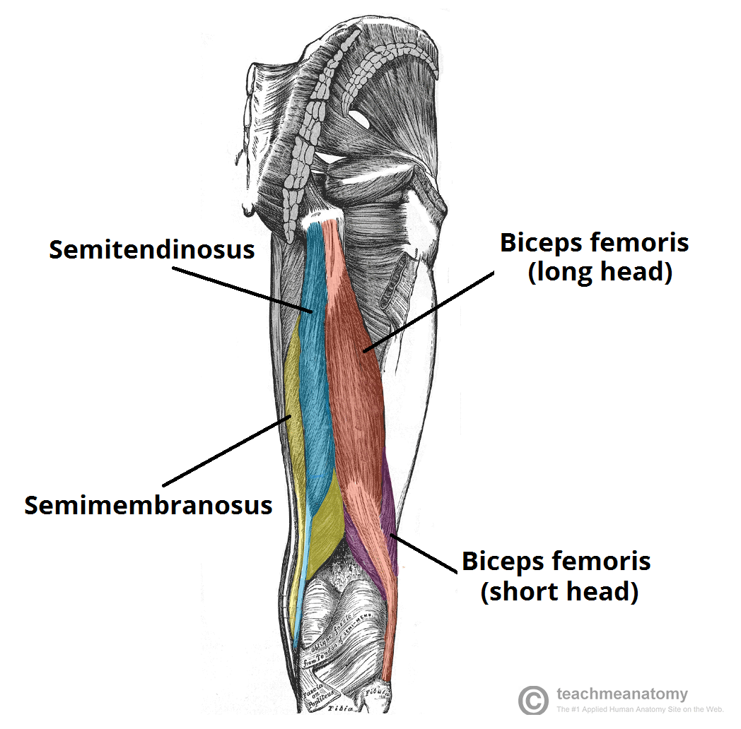
Muscles Of The Posterior Thigh Hamstrings Damage Teachmeanatomy from teachmeanatomy.info Medial muscles adduct and rotate your thigh, and posterior flex your leg and extend your thigh. Tendons are thick bands of tissue that connect muscles to bone. This is the group of muscles that you often see body builders flexing, which protrude just above the knee and take up most of the upper leg. Tendons are cords made of tough tissue, and they work as special connector pieces between bone and muscle. The large achilles tendon is the most important tendon for walking, running we created an anatomical atlas of the upper limb, an. The human leg, in the general word sense, is the entire lower limb of the human body, including the foot, thigh and even the hip or gluteal region. On the medial edge of the posterior thigh is the gracilis muscle. See more ideas about muscle anatomy, leg muscles anatomy, leg muscles.
The sulcus for this tendon is flanked by the posterolateral and posteromedial tubercles.
Related posts of muscle anatomy upper leg. The medial thigh muscles are responsible for the adduction (movement of a body part toward the body's midline) of the leg. This is why you have to indicate which biceps you are taking about when discussing one or other of these muscles. Related online courses on physioplus. Upper leg tendon anatomy : The hamstring muscles in the back of the thigh, the quadriceps muscles in the front, and the adductor muscles on the inside. It is also visible on the medial edge of the thigh from the anterior. The muscles of the leg anatomy chart shows in every possible view the way that the muscles and other pieces of the leg work together in motion and flexibility. It begins in the thigh area and extends to the head of the fibula in the knee. The vastus lateralis is a muscle located on the lateral, or outside, part of your thigh. On the medial edge of the posterior thigh is the gracilis muscle. Search photos retinaculum from t3.ftcdn.net 3d illustration back fit strong human anatomy. In clinical anatomy the thigh muscles are divided into three groups:
Tendons are cords made of tough tissue, and they work as special connector pieces between bone and muscle. The medial thigh muscles are responsible for the adduction (movement of a body part toward the body's midline) of the leg. Possibly the most important tendon in terms of mobility is the achilles tendon. It serves to attach the plantaris, gastrocnemius (calf) and soleus muscles to the calcaneus (heel) bone. The thigh has some of the body's largest muscles.
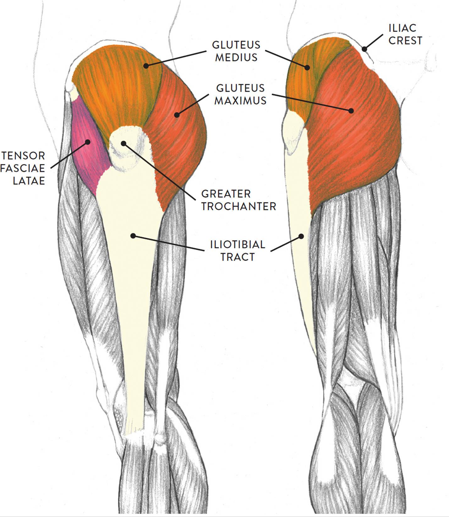
Muscles Of The Leg And Foot Classic Human Anatomy In Motion The Artist S Guide To The Dynamics Of Figure Drawing from doctorlib.info They are remarkably strong, having one of the highest tensile strengths found among soft tissues. This important tendon in the back of the calf and ankle connects the plantaris, gastrocnemius, and soleus muscles to. Tendons are thick bands of tissue that connect muscles to bone. This is why you have to indicate which biceps you are taking about when discussing one or other of these muscles. Notice the upper leg has a biceps muscle just like the upper arm does. See more ideas about muscle anatomy, leg muscles anatomy, leg muscles. They have a lot to do with how your hips move. Upper leg anatomy and function the upper leg is often called the thigh.
On the medial edge of the posterior thigh is the gracilis muscle.
The human leg, in the general word sense, is the entire lower limb of the human body, including the foot, thigh and even the hip or gluteal region. On the medial edge of the posterior thigh is the gracilis muscle. Possibly the most important tendon in terms of mobility is the achilles tendon. A muscle strain (muscle pull or tear) is a common injury, particularly among people who participate in sports. They consist of the rectus femoris, vastus intermedius, vastus lateralis and the vastus medialis. In clinical anatomy the thigh muscles are divided into three groups: Related online courses on physioplus. Rectus femoris these four muscles come together to form a single tendon, which inserts into the patella, or kneecap. Thigh muscles are responsible for allowing normal gait and proper lower extremity function (1). The thigh has three sets of strong muscles: Medial muscles adduct and rotate your thigh, and posterior flex your leg and extend your thigh. The muscles of the leg anatomy chart shows in every possible view the way that the muscles and other pieces of the leg work together in motion and flexibility. This is why you have to indicate which biceps you are taking about when discussing one or other of these muscles.
Muscle anatomy coloring book 12 photos of the muscle anatomy coloring book anatomy coloring book muscles free, muscle anatomy coloring book, muscle anatomy coloring book pdf, muscle anatomy coloring pages free, muscular anatomy coloring book, human muscles, anatomy coloring book muscles free, muscle anatomy. Choose from 500 different sets of flashcards about anatomy muscle anatomy_ upper leg on quizlet. The quadriceps tendon is located above the knee and attaches the. Its muscle belly is on the back aspect of the upper arm. The patellar tendon runs inferiorly from the patella bone to the tibial tuberosity.
Muscles Of The Leg And Foot from www.innerbody.com This important tendon in the back of the calf and ankle connects the plantaris, gastrocnemius, and soleus muscles to. The vastus lateralis is a muscle located on the lateral, or outside, part of your thigh. It also is active in maintaining thigh and kneecap position while walking and. We speak of the upper extremities (arms) and the lower extremities (legs). Squeeze your knees together and boom, you're contracting the adductors. Related online courses on physioplus. Related posts of muscles and tendons of the leg muscle anatomy coloring book. Other muscles of the anterior (front) thigh include the pectineus, sartorius,.
Your lower leg includes three main muscles, located behind your tibia or shinbone.
The quadriceps tendon attaches the quadriceps muscles to the patella. The medial thigh muscles are responsible for the adduction (movement of a body part toward the body's midline) of the leg. They have a lot to do with how your hips move. Lateral (fibular) collateral ligament (fcl) upper part middle part lower part popliteus tendon (pt) upper part i. Its muscle belly is on the back aspect of the upper arm. Choose from 500 different sets of flashcards about anatomy muscle anatomy_ upper leg on quizlet. Related posts of muscle anatomy upper leg. These muscles run from the lower spine. Upper leg tendon anatomy : Notice the upper leg has a biceps muscle just like the upper arm does. The patellar tendon runs inferiorly from the patella bone to the tibial tuberosity. Anterior muscles extend your legs and flex your thighs. On the medial edge of the posterior thigh is the gracilis muscle.